The Astrocyte Hypothesis
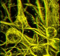
 Hat tip to the Bioethics blog for pointing out an article in the newest issue of Nature Neuroscience in which glial cells are shown to be capable of increasing blood flow dependent on recent neural activity. Glial cells, also known as astrocytes, can increase bloodflow by 37% in less than 2 seconds; such activity suggests they may have a more important regulatory role in processes such as learning. This study is one of several that are beginning to establish a more important role for astrocytes in information processing than previously thought.
Glial cells outnumber neurons in the cortex by almost 10 to 1, but were previously thought to be only simple "support cells" and not involved in the details of learning and memory - a role traditionally assigned only to neurons. However, Takano, Tian, Peng, Lou, Libionka, Han and Nedergaard have used 2 photon microscopy and laser Doppler flowmetry in live, exposed cortex of adult mice to show that glial cells are the means by which neural activity is tied to local blood flow. This is important because fMRI, probably the dominant form of brain imaging in use today, is not capable of imaging neural activity directly, but instead reflects changes in blood flow. In a sense, fMRI images astrocytic as opposed to neuronal activity, although as shown by this study they are tightly coupled.
The exact mechanism by which astrocytes cause vasodilation is thought to be triggered by increases in extracellular glutamate, triggering cytoplasmic calcium release and thereby signalling the production of both COX-1 and arachidionic acid derivatives. These are then converted to vasodilators by vasoactive epoxyeicosatrienoic acids.
Related Posts:
Complexity and Biologically Accurate Cognitive Modeling


0 Comments:
Post a Comment
<< Home